
What is Enterobiasis?
Enterobiosis (enterobius vermicularis from Greek. Enteron – intestines, bios – life, from lat. Vermis – worm) – a parasitic disease of a person, characterized by intestinal lesions, itching around the anus and allergization of the body.
Causes of Enterobiasis
The causative agent of enterobiosis is pinworm – Enterobius vermicularus (Linnaeus, 1758; Leach, 1853).
The name Enterobius comes from the Greek enteron – intestines and bios – life, vermicularis – reduced from Latin – a worm living in the intestines of a worm.
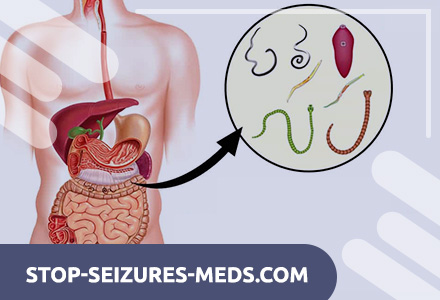
Pinworm is a small nematode of a spindle-shaped milk (milk) of white color, the cuticle of which has a transverse striated.
The length of the adult female reaches 9–12 mm, the male 3–5 mm, the tail end of the female is pointed, the male is blunt and hooked. Sharp lateral keels extending along the length of the helminth body form head vesicles at the anterior end. The helminth’s digestive system is represented by a mouth opening limited by three lips, a cylindrical esophagus with a bulbous extension extending into the intestine and ending in the anus in the back of the body.
The esophagus bulbus and vesicles form a suction device that secures the fixation of adult helminths to the intestinal wall.
The female reproductive system consists of a paired ovary, a uterus that passes into the vagina, and ends with a vulva. The vagina has muscle pulp, which is in a spasmodic state in the anoxic environment of the human intestine. Therefore, parasitic females, while in the intestinal lumen, do not secrete eggs. The male reproductive system is represented by the testis, which ends with a long spicule.
Pinworm eggs have an oblong, somewhat asymmetric shape, one side more flat. The size of the eggs is 50-60 x 20-30 microns. They are covered with a thin double-circuit colorless smooth shell.
Adult helminths live in the lower part of the small intestine, in the cecum and in the upper part of the colon. As a rule, only females parasitize, males after copulation are excreted with feces. Helminths feed on the contents of the intestines and are optional hematophages. The number of individuals parasitizing in the intestines varies widely from several tens to hundreds and thousands. K.I. Skryabin, V.P. Podyapolskaya and R.S. Schulz described a case when 2750 parasites were found in the intestines during autopsy of a child. Intensive infestations are associated with repeated self-infections.
Human infection with enterobiosis occurs when swallowing mature pinworm eggs, which contain motile larvae. Under the action of digestive enzymes of the small intestine, the larvae are released from the eggs, descending into its lower parts, 2-3 molts pass. Here the process of copulation is completed, and the males passively leave the intestines. And young female pinworms are attached to the mucous membrane with the help of head vesicles and the suction action of the bulb of the esophagus. Eggs form and accumulate in the uterus of the fertilized female, the number of which reaches 5-17 thousand. A stretched enlarged uterus squeezes the bulb of the esophagus, displaces it, as a result of which the helminth loses its ability to stay on the mucosa and, under the action of peristalsis, falls into the lower part of the large intestine. Further, during the active migration of helminth in the rectum, the eggs in the uterus ripen to the stage of a tadpole-like larva.
Overcoming the resistance of the sphincter of the rectum, the female creeps out onto the perianal folds and perineal skin of the invaded. The presence of atmospheric oxygen relaxes the genital tract of the helminth, as a result of which the creeping female secrete eggs that achieve invasiveness directly on the host’s body. As the female moves, heaps of eggs 100 to 300 in each remain on the skin of the invaded.
Pinworms creep out more often at night, when falling asleep and during sleep, when the muscles of the anus sphincter are somewhat weakened.
With considerable humidity and untidiness around the anus, oviposition is delayed, and pinworms continue to wander, sometimes crawling along the perineum not only into the vagina, but even through the uterus and fallopian tubes into the pelvic cavity, where they were found encapsulated on the peritoneum.
The individual life of the female who has secreted eggs ends, she dries, turning into a shapeless lump.
The itching that occurs with the movement of helminths leads to scratching of itchy places, contamination of the hands, getting eggs into the subungual beds, where the conditions for development to the invasive stage are also favorable.
Pathogenesis during Enterobiasis
The life cycle of the pinworm does not depend on the climatic conditions of the area, therefore enterobiasis is common in all latitudes of the world where humans exist, and a high level of infestation by pinworms is recorded in many countries.
The share of enterobiasis among other helminthiases reaches 70 – 95%. The spread of enterobiasis depends on the sanitary condition of the home, institution and personal hygiene skills of people. The invasion of children in preschool institutions and schools is especially high. There is no doubt the pathogenic effect of pinworms primarily on the child’s body, therefore enterobiasis is of great medical and social significance. It has been established that pinworms, exhibiting an immunosuppressive effect on the body, contribute to a more frequent occurrence in invasive somatic and infectious diseases.
The epidemiological significance of pinworms cannot be ignored either. For the first time ES Shulman pointed out the important role of pinworms in the spread of intestinal infections. It was found that the incidence rate of children with enterobiasis and AEI is in direct high correlation, and infested children are 2 – 3.5 times more likely to suffer from intestinal infections. This is natural, because the level of fecal contamination of the hands of infested children is 3.7 times higher than that of free from helminths. Enterobiasis also contributes to the transmission of other contact parasitoses – hymenolepiasis and giardiasis.
The only source of pinworm infestation is the infested person. The route of infection is oral. A person becomes infected by swallowing eggs. Fingers contaminated with pinworm eggs play an important role in the transmission of the invasion. Enterobiasis is correctly considered a disease of dirty hands. Due to the intolerable itching associated with the crawling of female pinworms from the rectum and their movement, the invasive contaminates the hands when scratching. With dirty hands, helminth eggs are carried onto other parts of the body – the skin of the abdomen, thighs, face, and also fall on underwear and bedding, and when shaken, they rise with dust and settle on household items, floor, furniture, creating conditions for the patient’s self-infection, – reinvasion and for widespread invasion of others.
When studying various objects of the external environment, pinworm eggs were found on dishes, toys, tables, desks, money, on food, where they fell from dirty hands and with dust.
Flies play a role in the spread of pinworm eggs.
On human skin, underwear, pinworm eggs ripen quickly (4-6 hours) to the invasive stage. The optimum temperature for their development is 35 – 37 ° С, but they are able to develop at temperatures from 23 to 40 ° С.
Helminth eggs at a temperature of 18 – 20 ° C and a relative humidity of 70% survive for 3 weeks, at -15 ° C for no more than 40 – 50 minutes, and at a temperature of + 55 ° C and above – a few seconds. Drying and solar radiation are detrimental.
The main factor in the transmission of infestation is hands, as well as toys, food contaminated with hands. Infestation of children in swimming pools is possible. The possibility of infection by inhaling eggs with dust is not excluded.
The pathogenic effect of pinworms on the human body is due to a complex of mechanisms.
The pathogenesis is based on the mechanical effect of helminths on the intestinal mucosa, associated with irritation of mechanoreceptors and chemoreceptors during their fixation and movement. Irritation of the ileocecal region leads to reflex disturbance of the motor and secretory functions of the digestive tract organs, and as a consequence – the possibility of developing gastritis, gastroduodenitis, enteritis.
Individual individuals of female helminths can penetrate deep into the mucous membrane 2/3 of their body length, as a result of which an inflammatory reaction develops around them, granulomas are formed, consisting of eosinophils, lymphocytes, macrophages. The severity of the pathological process depends on the intensity and duration of the invasion.
When entering the appendix, pinworms can cause appendicitis. Gutsaliev A.G. (1966) found pinworms in the appendix in 111 out of 322 operated on for appendicitis. In those infested with pinworms, anal and rectal fistulas, paraproctitis are described. Due to prolonged irritation and scratching on the skin of the perianal region, dermatitis and even severe weeping and dry eczema occur, sometimes extending far beyond the perineum.
Ectopic migration of pinworms is the cause of vulvitis, vaginitis, endometritis, constituting a serious pathology in pediatric gynecology.
Severe irritation of the perianal and genitourinary spheres and scratching with fingers lead to early awakening of the sexual instinct, to masturbation, masturbation, and dysuria.
In the pathogenesis of enterobiasis, as has been established in recent years, the immunosuppressive effect of pinworms is of great importance. According to A. Ya. Lysenko, Feldman, 1991; Markina, 1994, and others, pinworms suppress the development of post-vaccination immunity against diphtheria. Given the widespread occurrence of enterobiasis in children, the authors explain this by the high percentage of schoolchildren, preschoolers and disorganized children who are not immune to diphtheria.
Even after three times DPT inoculations, 18% of children lack anti-diphtheria antibodies, and 14.5% acquire them in very low titers. In connection with these data, an important conclusion is made that in order to achieve a high level of effectiveness of vaccination against diphtheria, it is necessary to free children from invasion before it is carried out.
Pinworms have a negative effect on the nervous system, especially children, their behavior, intelligence, causing increased irritability, absent-mindedness, impairment of memorization, etc.
The presence of pinworms leads to disruption of the normal composition of the intestinal microbial flora. Attention is drawn to the fact that dysbiosis and the prevalence of pathogenic microbial flora in the large intestine in the presence of invasion are recorded 4.5 times more often when compared with non-invasive children.
According to numerous observations, the level of morbidity in children with enterobiasis and acute intestinal infections is in a direct high correlation relationship; infested children are 2.5 – 3 times more likely to suffer from acute intestinal infections. Enterobiasis also adversely affects the course of intestinal infections of viral hepatitis A, in children – childhood infections.
Many observations confirm the aggravating effect of enterobiasis, like other helminthiases, on the course of pregnancy, causing toxicosis, dermatoses and other pathologies. Given the high contagiousness of the invasion, one cannot fail to foresee a third of its significance in terms of possible infection of newborns in the postpartum period, lactation period.
Symptoms of Enterobiasis
The clinical manifestations of enterobiasis are varied and depend on the intensity of infection, the frequency of reinvasion and the individual reaction of the infected person. In addition, numerous observations confirm the importance of age, namely, a more severe, often complicated reaction of the child’s body.
Observations of the parasitologist A.S. Kozlov (1985), who conducted a self-experiment, repeating pinworm infections seven times over the course of two years, are of indisputable interest. Achieving practical reliability, he swallowed different doses of eggs – 4 re-invasions and 2 super-invasions – from 1000 to 10 000. As a result, the author had reason to conclude that in the clinical course of enterobiasis, like other helminthiasis, there are two phases – acute and chronic …
In the case of primary invasion, according to these data, the clinical incubation is two days, the acute phase is 5-7. Parasitological incubation – the beginning of the isolation of females, which actually determines the duration of the chronic phase of one infection, is 35 – 75 days.
The main clinical symptoms of the acute phase of enterobiasis are abdominal pain, morning intestinal discomfort, nausea, and frequent bowel movements up to 4 times a day. These phenomena last up to 5 – 7 days, the stool remains speeded up until the end of the invasion.
The nature of clinical manifestations in the chronic phase is ambiguous. In some of the infected, enterobiasis proceeds without complaints, in the absence of clinical symptoms. The intensity of symptoms in the chronic phase of enterobiasis depends primarily on the intensity of invasion and the frequency of super- and reinvasion.
With a weak invasion, the main complaint is perianal itching, which occurs more often in the evening before bedtime, when falling asleep, and sometimes abdominal pain. After 2-3 days, all these symptoms disappear and, as a rule, recur every 3-4 weeks. This periodicity is associated with the completion of the development of a new generation of helminths as a result of reinvasion.
With a more intense and prolonged invasion, perianal itching disturbs the invader not only in the evening and night hours, but also during the day. Abdominal pain intensifies, more often in the right ileocecal region, reminiscent of pain in appendicitis, tenesmus, flatulence, and loose stools occur. Unbearable itching disrupts sleep, becomes the cause of nocturia, masturbation. From the side of the hemogram, moderate leukocytosis is noted, eosinophilia of peripheral blood, which gradually increases in the early phase, reaches a maximum (23%) by 16 days, then gradually decreases by the end of the invasion to 4 – 5%.
As a rule, neurological symptoms are expressed, such as headache, dizziness, memory loss. Children often suffer, they become capricious, whiny, get tired quickly, and do not perceive useful skills well. Instability of attention, inability to concentrate reduces academic performance, in adults – work capacity.
Enterobiasis has a reliably negative effect on behavioral reactions and neuropsychic development of preschool children. Cases of fainting and epileptiform seizures in children, causally associated with enterobiasis, have been described.
Severe complications of enterobiasis include appendicopathies and appendicitis, proctitis and paraproctitis, dermatitis and eczema of the perianal region with their characteristic symptoms.
The indisputable cause of painful vulvitis and vulvovaginitis in girls is often chronic enterobiasis.
M.G. Makarova found that enterobiasis causes a significant decrease in nonspecific immunity: the activity of salivary lysozyme (1.5 times) and the content of alpha-interferon (to a level less than 2 units / ml), at the same time does not affect the content gamma interferon. These data are of particular interest in terms of establishing the causes of immunosuppression, which is especially common in children.
The prognosis in the absence of complications with enterobiasis is favorable.
In conclusion, it is necessary to pay attention to the polymorphism of clinical manifestations causally associated with enterobiasis, but characteristic of the most diverse organ pathology. This is what leads to the inevitable appeal of the invasive to doctors of different specialties: gastroenterologists, obstetricians-gynecologists, neuropathologists, allergists, dermatologists, urologists, surgeons, etc. And only the correct orientation of such a specialist on the need to exclude enterobiotic invasion provides qualified assistance to the patient.